

The stochastic optical reconstruction microscope STORM technology can provide 10 times the resolution of traditional light microscopes, which is a powerful tool for observing the structure and localization of organelles and even biological macromolecules.
01 Introduction to the study

In 2020, researchers at the Biomedical Optics Imaging Laboratory at the University of Pittsburgh in the United States optimized the STORM imaging method and applied it to the observation of heterochromatin structures in traditional pathological paraffin sections of mouse intestinal epithelium, obtaining high-quality super-resolution images. The results revealed a hallmark feature of the early malignant transformation of tumor cells – the high-order folded structure of chromatin gradually becomes loose and fragmented. This may have a positive effect on improving the diagnosis, grading and prevention of cancer.
02Research content (excerpt)
Many studies have shown that defects in heterochromatin structures can lead to genomic instability and increase the risk of tumorigenesis. The authors used the optimized STORM imaging technique to observe the heterochromosomal structure labeled by H3K9me3, combined with a variety of measurement and analysis methods, and found that in normal tissues, the heterochromosomes formed large nanoclusters with high polymerization, while in the tumor model mice, the early heterochromosomal polymeric structure of carcinogenesis gradually spread, suggesting the disintegration of the heterochromatin structure.
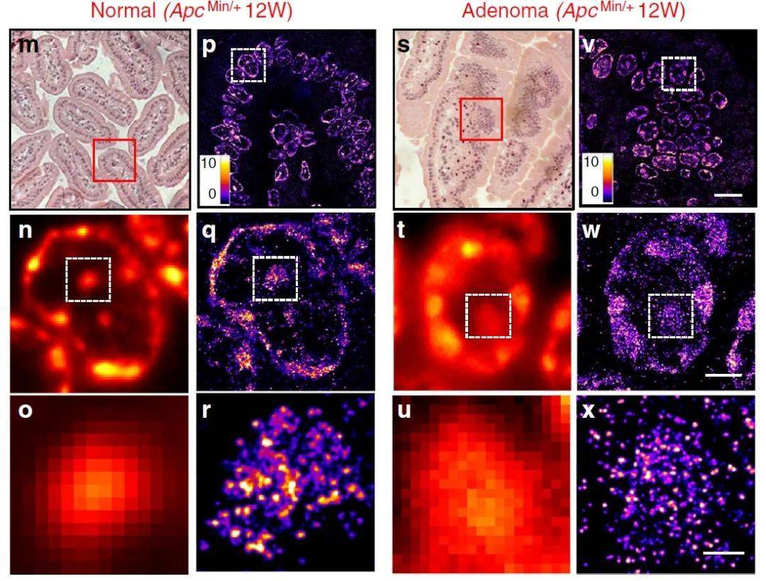
Figure 1: Super-resolution imaging of heterochromatin structure in ApcMin/+ mouse model.
The authors simultaneously took 3D-SIM images of DAPI staining of intestinal epithelial tissue of wild-type and tumor model mice, and measured the 3D chromatin structure, which showed that the fluorescence intensity and volume diffusion of the heterochromatin region decreased compared with normal tissue, which jointly suggested the disintegration of the heterochromatin structure.
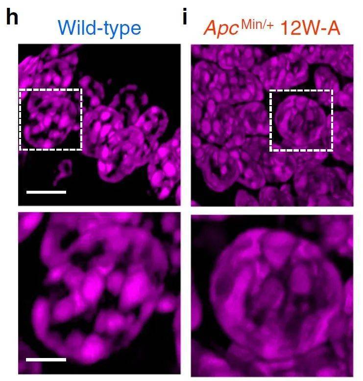
Figure 2, 3D-SIM image of DAPI-stained DNA folding in wild-type mouse normal cells and 12-week ApcMin/+ mouse tumor cells.
To investigate the effects of heterochromatin structural disruption on gene stability and transcription, the authors selected DNA double-strand break markers γ-H2AX sites as observation objects and found a significant increase in the number of γ-H2AX sites in specimens from tumor model mice, indicating DNA damage or genomic instability. The authors further observed the changes of the active transcription factor RNAPII during carcinogenesis and the colocalization relationship between small DNA nanoclusters and RNAPII after disintegration, demonstrating that transcriptional activity gradually increases during tumorigenesis.

Figure 3: Effects of chromatin structure disruption on transcription and genome stability.

Figure 4: Functions and structural consequences of heterochromatin structural disruption.
Based on the results of the study, the authors propose a molecular-scale model describing the structure of heterochromatin during carcinogenesis (Figure 5). That is, normal cells transform into tumor cells during carcinogenesis, and chromatin folding is gradually decompressed and fragmented, with heterochromatin focus enlargement, thereby enhancing transcription factory formation and genomic instability.
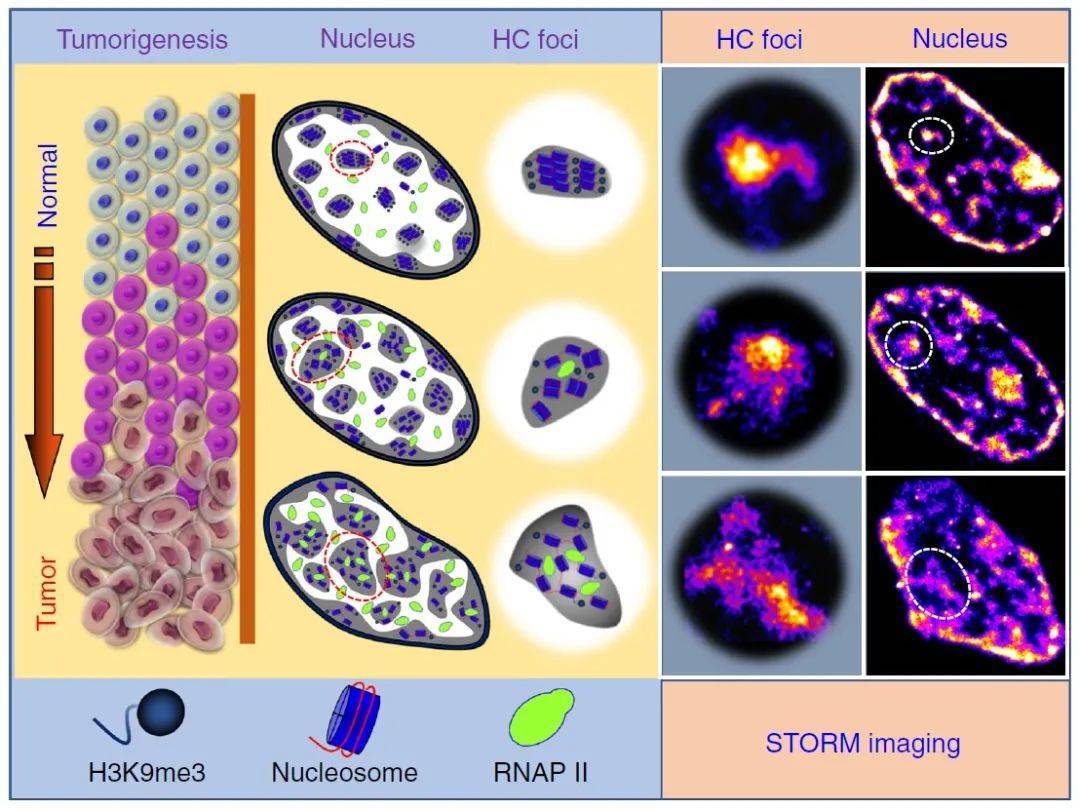
Figure 5: A molecular-scale model describing the structure of heterochromatin during carcinogenesis.
03 Ultra-high resolution microscopy imaging system iSTORM appointment test shoot
The released super-resolution microscopic imaging system iSTORM has successfully achieved a breakthrough in the diffraction limit of optical microscopy, making it possible to engage in single-molecule localization and counting of biological macromolecules, subcellular and supramolecular structure analysis, and biodynamics of biological macromolecules at a resolution scale of 20 nm, thus bringing major breakthroughs to life sciences, medicine and other fields.

The super-resolution microscopy imaging system iSTORM has the characteristics of 20 nm ultra-high resolution, 3-channel simultaneous imaging, 3D simultaneous shooting, real-time reconstruction, and 2-hour novice mastery, and has realized the localization and counting of single molecules in live cells, and provides fluorescent dye selection, sample preparation, imaging services and experimental solutions as an overall solution. It has been highly recognized by more than 50 scientific research groups and more than 100 researchers.
Reference links:
https://www.nature.com/articles/s41467-020-15718-7