

Recently, Professor Yang Liu's team at the University of Pittsburgh has made important progress in super-resolution microscopic imaging of patient tissue chromatin structure, the team has designed a small molecule probe that can be directly used for super-resolution imaging of patient tissue DNA structure, and used random optical reconstruction microscopy (STORM) to achieve simple, reliable and rapid high-resolution imaging of patient tissue chromatin structure, which is of great significance for studying the role of chromatin structure in the process of carcinogenesis. The relevant results were published in the authoritative journal Science Advances, and Dr. Jianquan Xu is the first author of the article.
01Research Introduction (Excerpt)
It is well known that at the microscopic scale, changes in the structure of the nucleus or chromatin are one of the most typical characteristics of cancer cells, and it is also an important basis for pathologists to detect cancer or determine its phenotype under light microscopy. However, current research does not know how chromatin changes at the molecular scale during carcinogenesis.
Due to the limitation of resolution and the optical diffraction limit, changes in chromatin at the molecular level cannot be observed with conventional light microscopy. In recent years, the rapid development of super-resolution fluorescence microscopy technology has broken the limitation of traditional light microscopy on resolution, increasing the resolution to tens or even several nanometers, greatly expanding our ability to study biomolecules and biological processes at the cellular and subcellular levels.
Among the various super-resolution microscopy techniques, stochastic optical reconstruction microscopy (STORM) works relatively simply and uses the photoconversion properties of fluorescent molecules to obtain superior spatial resolution, so it is widely used. It is of great significance to apply this technology to observe the microstructural changes of the nucleus during carcinogenesis, so as to study the pathogenesis of cancer and provide more accurate cancer detection standards.
Formalin-fixed paraffin-embedded tissue (FFPE) is the most commonly used specimen in medicine, and the fluorescent molecules currently routinely used for STORM imaging cannot be used for super-resolution imaging of chromatin in paraffin-embedded tissue, or cannot achieve high resolution, which greatly affects the application of this technology in this field.
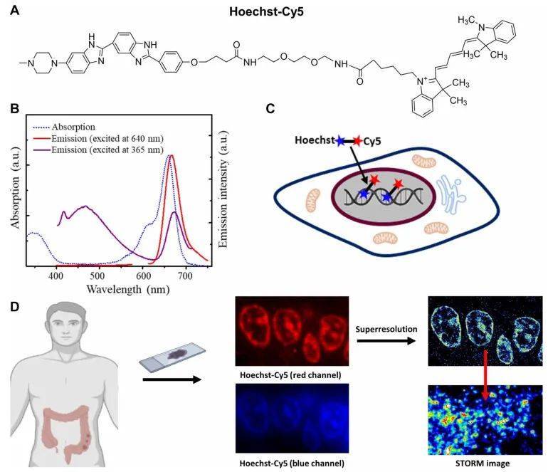
Liu Yang's research team at the University of Pittsburgh has coupled the Cyanine-5 molecule with superior photoconversion properties with the DNA stain Hoechst (Hoechst-Cy5) to achieve rapid staining of DNA in FFPE tissues, and uses the photoconversion properties of Cyanine-5 for high-resolution STORM imaging, thereby visualizing the ultrastructure of chromatin in FFPE tissue cells, and successfully applied this technology to the detection of patient samples.
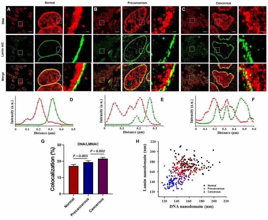
Through STORM super-resolution imaging of DNA in the nucleus of multiple groups of patient PPFE samples, they found that intranuclear chromatin became looser and nucleosome clusters became smaller during carcinogenesis. The looser structure of this chromatin is closely related to many functions of the cell, such as the transcription process of the cell and the structural changes of the nuclear fiber layer.
Using two-color STORM imaging, the researchers found that the more loose chromatin in cancer cells promoted the transcriptional activity of the cells, manifested by increased size of RNA polymerase (RNAPII) clusters and increased colocalization with loosely structured chromatin. In addition, during the process of carcinogenesis, the structure of the nuclear membrane changes significantly, the chromatin loosening at the nuclear membrane is more pronounced, and the structure of the nuclear nuclear fiber layer is destroyed. This change may be related to the function of lamina-associated domains (LADs) in the process of carcinogenesis.
The researchers further applied this technique to detect tissue samples from patients with Lynch syndrome. The experimental results showed that cells in patients with Lynch syndrome had a looser chromatin structure than healthy people, which may indicate a higher risk of cancer. This finding may have significant implications for risk assessment and early diagnosis of cancer.
02 Super-resolution microscopic imaging system iSTORM scheduled test shooting
The STORM imaging technology mentioned above has been successfully commercialized, and experts and teachers who need STORM imaging technology for experimental research can fill in the questionnaire at the end of the article to make an appointment to get the iSTORM ultra-high resolution microscopy imaging system test shooting service~
iSTORM, the super-resolution microscopic imaging system released by Lixian Intelligence, has successfully achieved a breakthrough in the diffraction limit of optical microscopy, making it possible to engage in single-molecule localization and counting of biological macromolecules, subcellular and supramolecular structure analysis, and biodynamics of biological macromolecules at a resolution scale of 20 nm, thus bringing major breakthroughs to life sciences, medicine and other fields.
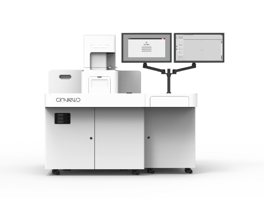
The super-resolution microscopy imaging system iSTORM has the characteristics of 20 nm ultra-high resolution, 3-channel simultaneous imaging, 3D simultaneous shooting, real-time reconstruction, and 2-hour novice mastery, and has realized the localization and counting of single molecules in live cells, and provides fluorescent dye selection, sample preparation, imaging services and experimental solutions as an overall solution. It has been highly recognized by more than 50 scientific research groups and more than 100 researchers. Welcome to book a test shoot!
References:
Ultrastructural visualization of chromatin in cancer pathogenesis using a simple small-molecule fluorescent probe ,Jianquan Xu, Xuejiao Sun, Kwangho Kim, Rhonda M. Brand, Douglas Hartman, Hongqiang Ma, Randall E. Brand, Mingfeng Bai, Yang Liu,Sci. Adv., 2022, DOI: 10.1126/sciadv.abm8293