

"There is still a river under the cell membrane city, what should I do?"
MLKL molecules are in a hurry,
Suddenly, I saw wooden blocks on the river.
Groups of more than four people,
Step on a raft made of more than four wooden blocks,
You can have the opportunity to cross the river.
Come to the city of cell membranes...
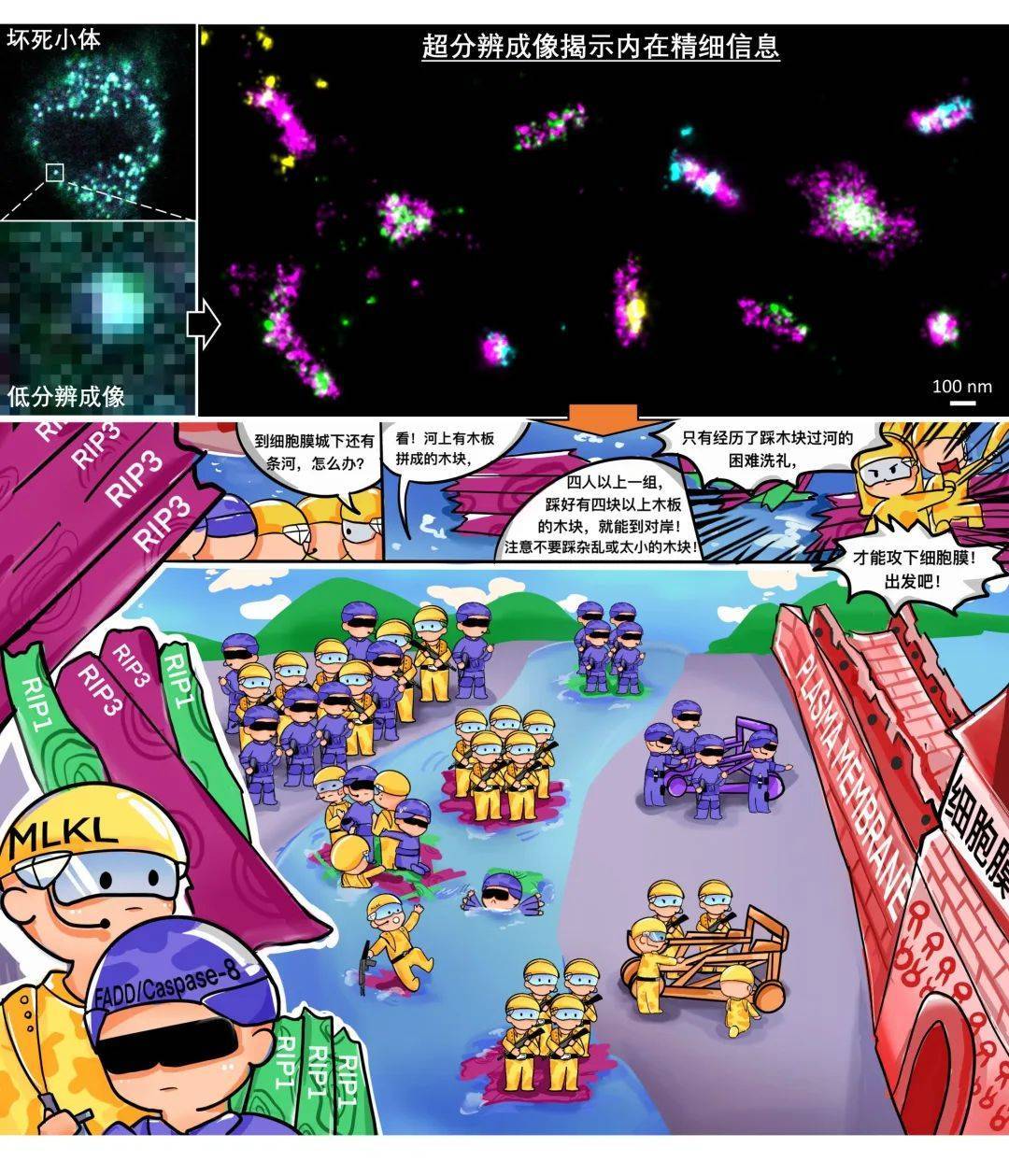
This year, the team of Han Jiahuai, academician of the Chinese Academy of Sciences and professor of Xiamen University, and Chen Xin, associate professor of Xiamen University, used the single-molecule localization super-resolution imaging technology "random optical reconstruction microscope (STORM)" to reveal for the first time the organizational structure characteristics of "necrotic bodies" in cells and their decisive role in cell death, providing new ideas for the treatment and intervention of human related diseases. The paper has been published in Nature Cell Biology.

01Ultra-high-resolution imaging technology makes inferences believable
As we all know, cells are the basic functional units of living organisms, and although they are very small in size, they have irreplaceable significance for living organisms. The key to determining the fate of cells is the programmed death of cells. In programmed cell death, there is a form called "necrotic apoptosis", and an important signal processing hub that plays a decisive role is the "necrostosome" complex.
What exactly is the structure of "necrotic bodies" in dead cells? How does "necrotic body" accurately determine the fate of cell death? These problems involve complex processes such as recruitment, activation, and signal amplification/transition of multiple core molecules (RIP1/RIP3/MLKL). Because the size of the cell body is very small, for example, mammalian cells are generally tens of microns, it is difficult to observe the precise regulatory mechanism of its internal "necrotic bodies".
In previous studies, scientists have used conventional confocal fluorescence microscopy to observe that cell death processes produce dot-like signals of different sizes of "necrossomes", suggesting that the signal hub is likely to have a dynamic assembly process. But how "necrossomes" accurately process complex signals in cells and determine cell death is always an unsolved mystery.
With the help of super-resolution imaging technology that has developed rapidly in recent years, the teams of Han Jiahuai and Chen Xin have tried a variety of currently mature technical schools, and finally found a weapon to accurately observe the operation mechanism of "necrosoids" - single-molecule localization super-resolution imaging technology (STORM).
By meticulously optimizing the whole process of STORM imaging, the team achieved more than 10 times the resolution (13~18nm positioning accuracy) on biological samples that was better than that of conventional confocal microscopes. The improvement of these technologies has made many research objects that were originally invisible and invisible become clear and clear, so that the conclusions originally obtained by speculation are "seen is believed".
02 Stochastic optical reconstruction microscopy (STORM) discovers how necrotic bodies accurately process complex signals
Returning to the above comic schematic, the team of Han Jiahuai and Chen Xin successfully observed that the "necrosomes" in dead cells evolved from the initial point-like mass structure to a regular rod-like structure with a diameter of about 50 nm and a length of about 200~600 nm, and showed an obvious mosaic-like distribution composed of RIP1/RIP3 in the regular rod-like structure. When MLKL groups of four people and finds more than four RIP3 wooden blocks, they can cross the river of "necrotic bodies" and then target the cell membrane, resulting in cell death.
"This result reveals in situ the tissue properties of key signaling hubs at the nanoscale and their contribution to signal transmission/amplification/transformation, providing a potential entry point for the development of interventions that specifically inhibit programmed cell death, and hopes that our findings can be helpful for the clinical response and treatment of neurodegenerative diseases (Parkinson's disease, multiple sclerosis, etc.) and pathogenic bacterial infectious diseases (sepsis, etc.)." Academician Han Jiahuai introduced.
03Ultra-high-resolution imaging technology is expected to resolve more biomacromolecular complexes
A large number of biological macromolecular complexes in cells are core functional hubs that control life activities, such as DNA replication/transcription initiation complexes and various transport complexes on organelle membranes. It has been nearly two hundred years since the birth of the cell theory, the cornerstone of modern biology, but human beings have never been able to completely analyze the fine molecular structure of any cell under homeostasis/stress conditions, and naturally cannot modify/control cells at will to achieve the grand goal of ensuring human health and social progress.
At present, single-particle cryo-EM technology is a powerful tool for analyzing protein structure, but it still has obvious limitations in the face of functional complexes with huge intracellular structure, complex composition and high heterogeneity. The work of Han Jiahuai and Chen Xin's team proves that nanoscale optical imaging is one of the feasible solutions to analyze the organizational characteristics and functional patterns of such large biomacromolecular complexes.
"The successful observation of the fine structure of cells in situ 'necrosomes' undoubtedly increases our confidence in using super-resolution imaging technology to reveal more of the internal laws of life in the future." Chen Xin said that in the future, with the further development of cutting-edge imaging technologies, such as single-nanometer resolution precision microscopy, cryotomography technology, etc., human beings will one day be able to clearly observe the magical inner laws of life that are always in operation.
04 Super-resolution microscopy imaging system iSTORM scheduled a test shot
The STORM imaging technology mentioned above has been successfully commercialized, and experts and teachers who need STORM imaging technology for experimental research can make an appointment for trial shooting. iSTORM, the super-resolution microscopic imaging system released by Lixian Intelligence, has successfully achieved a breakthrough in the diffraction limit of optical microscopy, making it possible to engage in single-molecule localization and counting of biological macromolecules, subcellular and supramolecular structure analysis, and biodynamics of biological macromolecules at a resolution scale of 20 nm, thus bringing major breakthroughs to life sciences, medicine and other fields.

The super-resolution microscopy imaging system iSTORM has the characteristics of 20 nm ultra-high resolution, 3-channel simultaneous imaging, 3D simultaneous shooting, real-time reconstruction, and 2-hour novice mastery, and has realized the localization and counting of single molecules in live cells, and provides fluorescent dye selection, sample preparation, imaging services and experimental solutions as an overall solution. It has been highly recognized by more than 50 scientific research groups and more than 100 researchers.
Paper Link:
https://www.nature.com/articles/s41556-022-00854-7
*Part of the content is from China Biotechnology Network, and the infringement contact is deleted