
01 Introduction to the study
Histological analysis of brain tissue samples gives us valuable information about the pathological processes that lead to common neurodegenerative diseases. In this context, the development of new high-resolution imaging methods is a current challenge in neuroscience. To do this, we use a super-resolution imaging technique called stochastic optical reconstruction microscopy (STORM) to analyze human brain slices. The authors combined the STORM cell imaging protocol with neuropathological techniques to image brain samples from patients and control subjects with neurodegenerative diseases.
02Research Results (Excerpt)
The authors performed 2D, 3D, and bicolor STORM imaging in neocortical, white matter, and brainstem samples. STORM proved particularly effective in visualizing the organization of dense protein inclusions, and the authors performed on pathological aggregates within the central nervous system of patients with Alzheimer's disease, Parkinson's disease, dementia with Lewy bodies, and frontotemporal degeneration Imaging < 50 nm resolution. Aggregated AB branches are reticulated and crosslinked in the extracellular matrix with widths ranging from 60 to 240 nm. Neuronal Tau and TDP-43 inclusions are denser, with celloids honeycomb-shaped and axons in filamentous tissue. Finally, STORM imaging of α-synuclein pathology revealed the internal organization of Lewy bodies, which cannot be observed by conventional fluorescence microscopy.
1. Imaging of frozen samples of the prefrontal cortex of the human brain using STORM and TEM measurements
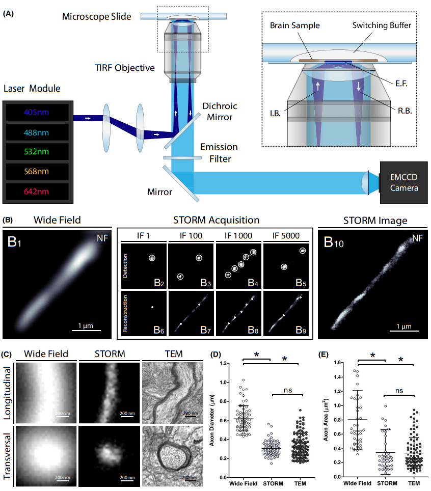
Figure 1: Super-resolution imaging of human brain samples using STORM.
(A) Schematic of the optical setup for STORM imaging. I.B., incident beam; E.F., asympathetic field; R.B., reflected beam.
(B) STORM acquires cortical axons in human brain slices for immunostaining of neurofilaments (NF): traditional wide-field fluorescence microscopy images are acquired first.
(B1), then strongly increase the excitation power to induce fluorophore scintillation and obtain thousands of frame recordings (B2-B5). The localization of activated fluorescent molecules is detected on a per-frame basis with subpixel precision (B6-B9). The super-resolution image (B10) is then reconstructed using cumulative positioning from all frames. IF, imaging frame.
(C) Representative images of longitudinal and transverse sections of prefrontal cortical axons obtained using conventional wide-field fluorescence microscopy, STORM, and transmission electron microscopy (TEM).
(D and E) axon diameter (longitudinal section) and area (transverse section) measured in the human brain using conventional fluorescence microscopy, STORM, and TEM. Error bars represent mean values with standard deviation. *P < .001
2. STORM image of age plaques and neurofibrillary tangles in brain samples of AD patients
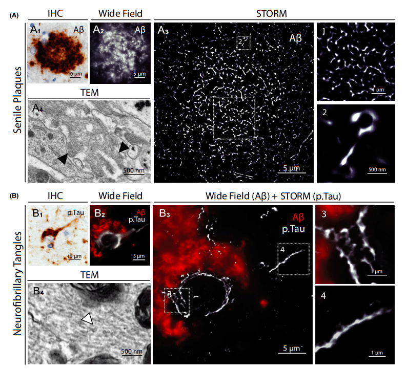
Figure 2: STORM image of age plaques and neurofibrillary tangles in brain samples from AD patients.
(A1) Representative image of age spots in neocortex in AD patients (immunohistochemical detection of Ab).
(A2) Immunostaining of AB with conventional fluorescence microscopy images of whole senile plaques in neocortical sections of the same patient. (A3) Storm image of the same area. The illustrations (1 and 2) show close-up details of the distribution and size of the aggregated AB branches. (A4) Comparative TEM image of Ab fibers (black arrows) in age spots.
(B1) Representative images of neurofibrillary tangles in neocortex in AD patients (P. Immunohistochemical detection of Tau).
(B2) Conventional fluorescence microscopy images of intracellular neurofibrillary tangles of entire degenerative neurons surrounded by AB deposition in neocortical sections of the same patient.
(B3) Imaging of the same neuron by combining conventional fluorescence microscopy (Ab) and STORM (p.Tau). Illustrations (3 and 4) show close-up details of the honeycomb structure of p. Tau aggregates in the soma and filamentous tissue in axons.
(B4) Comparative TEM image of Tau filaments (white arrows) in neurofibrillary tangles.
03 Research Summary
In this paper, the authors combine super-resolution microscopy and neuropathological techniques to analyze human brain slices. To date, imaging nanostructures in tissues has relied primarily on transmission electron microscopy, a time-consuming technique that requires rigorous sample preparation with ultrathin tissue sections (50-70 nm) and limits immunotargeted diversity and 3D acquisition. In contrast, STORM has the advantages of optical fluorescence microscopy in sample preparation, vast observation fields, multimolecular markers, and 3D acquisition, while image acquisition and reconstruction takes only a few minutes. STORM imaging of human brain samples further opens the door to a comprehensive understanding of common neurological disorders. The convenience of this technique should directly expand its application in super-resolution imaging of the human brain, providing better solutions to the current challenges facing neuroscience.
04 Super-resolution microscopic imaging system iSTORM
The random optical reconstruction microscope (STORM) technology mentioned above has been successfully commercialized, and experts and teachers who need STORM technology for experimental research, please fill in the questionnaire at the end of the article to make an appointment to get the iSTORM super-resolution microscopy imaging system test shooting service~
The super-resolution microscopy imaging system iSTORM has successfully achieved a breakthrough in the diffraction limit of optical microscopy, making it possible to engage in single-molecule localization and counting of biological macromolecules, subcellular and supramolecular structure analysis, and biodynamics of biological macromolecules on a resolution scale of 20 nm, thus bringing major breakthroughs to life sciences, medicine and other fields.
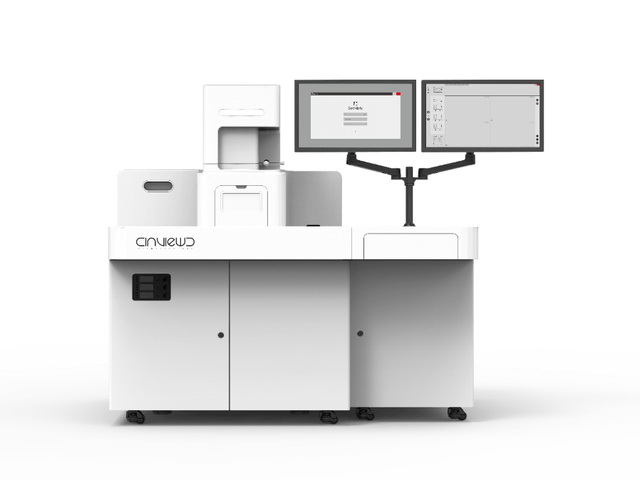
Figure 3: iSTORM, a super-resolution microscopy imaging system.
Ultra-high resolution microscopy imaging system iSTORM has the characteristics of 20 nm ultra-high resolution, 3-channel simultaneous imaging, 3D simultaneous shooting, real-time reconstruction, and 2-hour novice mastery, etc., has realized the localization and counting of single molecules in living cells, and provides fluorescent dye selection, sample preparation, imaging services and experimental solutions as an overall solution. It has been highly recognized by more than 50 scientific research groups and more than 100 researchers.
References:
P. Codron, F. Letournel, S. Marty, L. Renaud, A. Bodin, M. Duchesne, C. Verny, G. Lenaers, C. Duyckaerts, J.-P. Julien, J. Cassereau and A. Chevrollier (2021) Neuropathology and Applied Neurobiology 47, 127–142 STochastic Optical Reconstruction Microscopy (STORM) reveals the nanoscale organization of pathological aggregates in human brain